Journal of
eISSN: 2572-8466


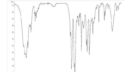
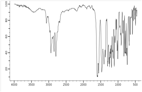
In the current study, we have experimentally and comparatively investigated and compared malignant human cancer cells and tissues before and after irradiating of synchrotron radiation using Atomic Force Microscopy Based Infrared (AFM–IR) Spectroscopy and Nuclear Resonance Vibrational Spectroscopy. It is clear that malignant human cancer cells and tissues have gradually transformed to benign human cancer cells and tissues under synchrotron radiation with the passage of time (Figures 1 & 2).1–145 It should be noted that malignant human cancer cells and tissues were exposed under white synchrotron radiation for 30 days. Furthermore, there is a shift of the spectrum in all of spectra after irradiating of synchrotron radiation that it is because of the malignant human cancer cells and tissues shrink post white synchrotron irradiation with the passage of time. In addition, all of the figures are related to the same human cancer cells and tissues. Moreover, in all of the figures y–axis shows intensity and also x–axis shows energy (keV) (Figures 1 & 2).101–145

A

B
Figure 1 Atomic force microscopy based infrared (AFM–IR) Spectroscopy analysis of malignant human cancer cells and tissues (A) before and (B) after irradiating of synchrotron radiation in transformation process to benign human cancer cells and tissues with the passage of time.1–145

A

B
Figure 2 Nuclear resonance vibrational spectroscopy analysis of malignant human cancer cells and tissues (A) before and (B) after irradiating of synchrotron radiation in transformation process to benign human cancer cells and tissues with the passage of time.1–145
It can be concluded that malignant human cancer cells and tissues have gradually transformed to benign human cancer cells and tissues under synchrotron radiation with the passage of time (Figures 1 & 2).1–145 It should be noted that malignant human cancer cells and tissues were exposed under white synchrotron radiation for 30 days. Furthermore, there is a shift of the spectrum in all of spectra after irradiating of synchrotron radiation that it is because of the malignant human cancer cells and tissues shrink post white synchrotron irradiation with the passage of time. In addition, all of the figures are related to the same human cancer cells and tissues. Moreover, in all of the figures y–axis shows intensity and also x–axis shows energy (keV) (Figures 1 & 2).101–145
None.
The authors declare that there is no conflict of interest.

© . This is an open access article distributed under the terms of the, which permits unrestricted use, distribution, and build upon your work non-commercially.